
A 31 years Right handed gentleman presented to my outpatient clinic with the history of pain over the left side of the chest for a week duration.The pain was not related to excretion or exercise and he had no symptoms suggestive of pleurisy.He could not recall any trauma or physical strain. He had no systemic symptoms such as fever.He had no significant past medical or surgical history of note.
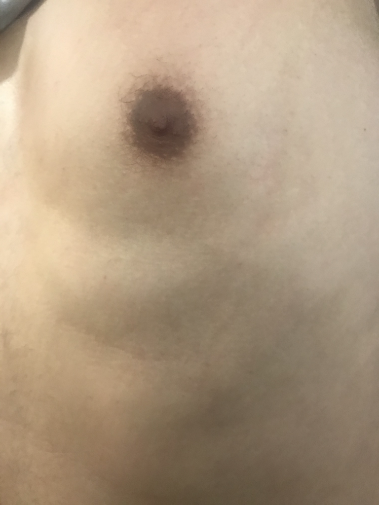
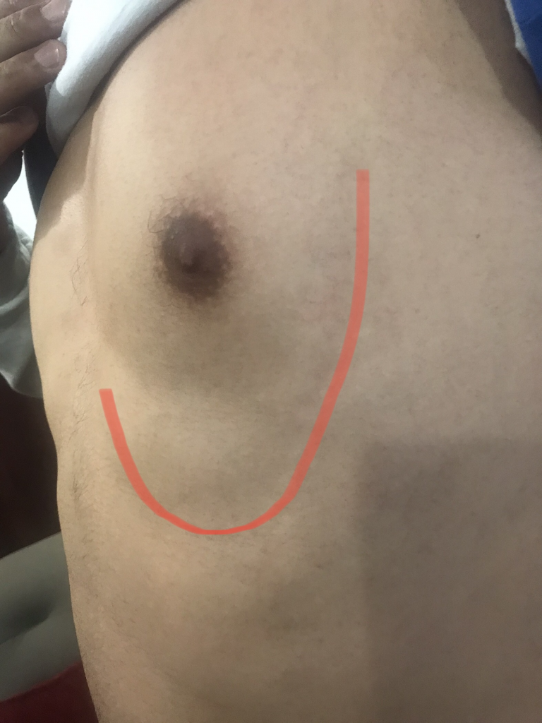
An examination revealed a tender area on left anterolateral part of his chest almost circling half to his Left sided breast with a cord-like swelling just under the skin extending from lateral aspect of left breast and inferior to the same breast, which was made prominent by abduction of his left arm.These finding were compatible with a clinical diagnosis of inflammation and thrombosis of some superficial vein. No other abnormalities were found.
USG showed prominent superficial vein with thrombus within the lumen, likely s/o superficial thrombophlebitis. Color doppler study shows no flow within the lumen.
Investigations to rule out hyper coagulable state were advised and he was sent home with NSAID.
Literature Review:
Mondor’s disease, as originally described by Mondor in 1939, is a superficial thrombophlebitis of anterior chest wall [1]. In 1958, Helm and Hodge reported a case of thrombosis of dorsal superficial vein of penis as penile Mondor’s disease [2]. Thoracoepigastric, superficial epigastric, and lateral thoracic veins are the most common sites of involvement. In our patient, the thoracoepigastric vein was involved. Although hormone therapy, breast cancer, thrombophilic conditions, and surgical or physical trauma, have been identified as the common etiological factors, mostly commonly it is idiopathic [3]. It is three times more common in females than in males [4].
Based on the history, clinical examination, and investigations, no risk factor was identified in this patient. Patients usually present with sudden onset of chest pain and a palpable tender erythematous cord-like structure is found corresponding to the anatomical location of the superficial veins of the chest wall or breast [5].
Even though the pathophysiology of this condition is not very clear, it is postulated that direct trauma, pressure on the vein with stagnation of blood, and stretching and relaxing of the vein are the underlying mechanisms for development of Mondor’s disease [6]. In an ultrasound scan, the thrombosed vessel appears as a superficially located, long, tubular, anechoic structure with a beaded appearance and absent color flow in Doppler studies [7]. In patients with palpable findings in the breast examination, mammographic examination is essential to rule out an underlying malignancy [7].
Although it is a self-limiting condition, local application of heat and topically or orally administered non-steroidal anti-inflammatory drugs can be used to alleviate the symptoms. Antibiotics are not necessary unless there is evidence of infection [8, 9]. Systemic anticoagulation with low molecular weight heparin or orally administered anticoagulants is recommended only for high-risk patients. Reduction in thrombus size and pain has been observed with locally acting anticoagulants, and it can be used in low-risk patients [10].
Chest pain is a common presentation encountered in clinical practice. Our patient presented with chest pain and was found to have the typical features on examination; diagnosis of Mondor’s disease was made based on the history and examination. No risk factor or underlying cause was identified in this case and diagnosis was confirmed further with duplex scan. Her symptoms resolved in 4 weeks with non-steroidal anti-inflammatory drugs. Mondor’s disease is a rare cause of chest pain which is often underdiagnosed due to lack of awareness. Failure to recognize this mostly benign self-limiting condition can result in unnecessary investigations and cause significant anxiety to the patient.
Ref:
1. Mondor H. Tronculite sous-cutané subaigue de laparoi thoracique antero-laterale. Mem Acad Chir (Paris) 1939;65:1271–8. [Google Scholar]2. Helm JD, Jr, Hodge IG. Thrombophlebitis of a dorsal vein of the penis: report of a case treated by phenylbutazone (Butazolidin) J Urol. 1958;79:306–7. doi: 10.1016/S0022-5347(17)66273-6. [PubMed] [CrossRef] [Google Scholar]3. Laroche JP, Galanaud J, Labau D, et al. Mondor’s disease: What’s new since 1939? Thromb Res. 2016;130:S56–8. doi: 10.1016/j.thromres.2012.08.276. [PubMed] [CrossRef] [Google Scholar]4. Viana GAP, Okano FM. Superficial thrombophlebitis (Mondor’s Disease) after breast augmentation surgery. Indian J Plast Surg. 2008;41(2):219–21. doi: 10.4103/0970-0358.44940. [PMC free article][PubMed] [CrossRef] [Google Scholar]5. Whitaker-Worth DL, Carlone V, Susser WS, Phelan N, Grant-Kels JM. Dermatologic diseases of the breast and nipple. J Am Acad Dermatol. 2000;43:733–51. doi: 10.1067/mjd.2000.109303. [PubMed] [CrossRef] [Google Scholar]6. Hogan GF. Mondor’s disease. Arch Intern Med. 1964;113:881–5. doi: 10.1001/archinte.1964.00280120081015. [PubMed] [CrossRef] [Google Scholar]7. Shetty MK, Watson AB. Mondor’s disease of the breast. Am J Roentgenol. 2001;177(4):893–6. doi: 10.2214/ajr.177.4.1770893. [PubMed] [CrossRef] [Google Scholar]8. Kikano GE, Caceres VM, Sebas JA. Superficial thrombophlebitis of the anterior chest (Mondor’s disease) J Fam Pract. 1991;33:643–4. [PubMed] [Google Scholar]9. Cesarone MR, Belcaro G, Agus G, et al. Management of superficial vein thrombosis and thrombophlebitis: Status and expert opinion document. Angiology. 2007;58:7S–14. doi: 10.1177/0003319706297643. [PubMed] [CrossRef] [Google Scholar]10. Cesarone MR, Belcaro G, Corsi M, et al. Local heparin, superficial vein thrombosis. Angiology. 2007;58:36S–41. doi: 10.1177/0003319706297647. [PubMed] [CrossRef] [Google Scholar]